Pelvic Anatomy Posterior : Pelvic Posterior View Of Ligaments Page 4 Line 17qq Com : Histologic analysis included that of 2 young nulliparous women whose tissue was harvested within 12 hours of death.
Pelvic Anatomy Posterior : Pelvic Posterior View Of Ligaments Page 4 Line 17qq Com : Histologic analysis included that of 2 young nulliparous women whose tissue was harvested within 12 hours of death.. This quiz is unlabeled so it will test your knowledge on how to identify these structural locations (iliac crest, ischial spine, acetabulum, superior ramus of pubis, posterior superior/inferior iliac spine, lessier. At times, it also may refer to structures or tissues found within or attaching to these bones. •two hip bones, which form the anterior and lateral walls. The pelvic region is the area between the trunk — or main body — and the lower extremities, or legs. When relaxed it works synchronously w/ the abdominal diaphragm absolutely necessary for efficient movement of lymphatic fluid away from pelvis and perineal tissues somatic dysfunction of pubic symphysis or innominates can place asymmetric tensions(on pelvic and urogenital diaphragm) pain:
It lies posterior to the uterus and anterior to the rectum. The pelvic region is the area between the trunk — or main body — and the lower extremities, or legs. The borders of the pelvic inlet: The pelvic bones are smaller and narrower. Bones of the pelvis and lower back the bones of the pelvis and lower back work together to support the body's weight, anchor the abdominal and hip muscles, and protect the delicate vital organs of the vertebral and abdominopelvic cavities.
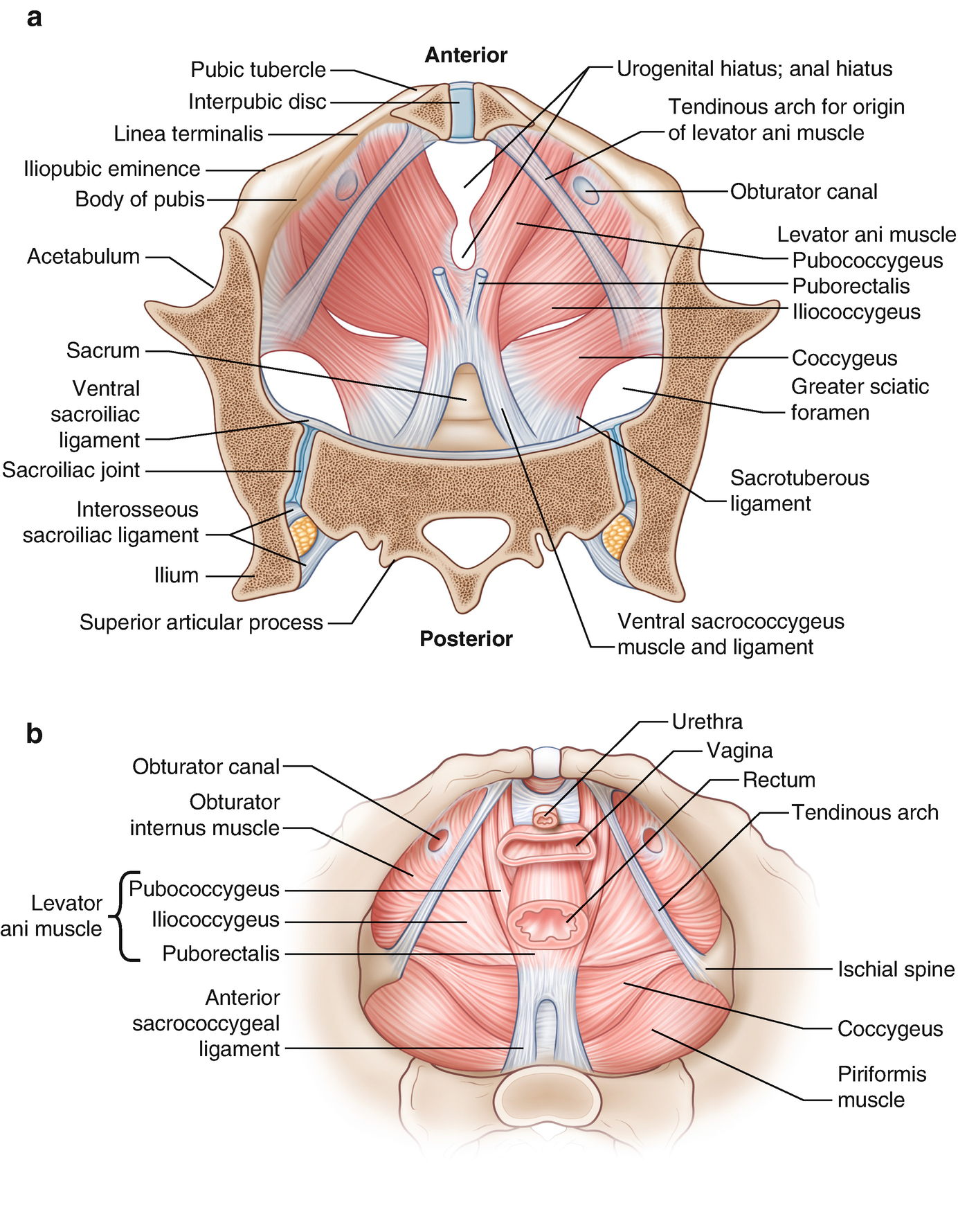
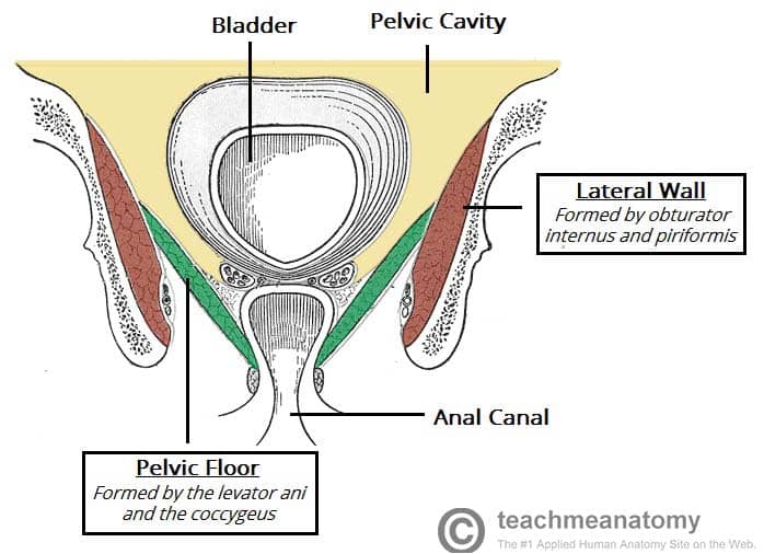
Muscles Of The Pelvic Floor Anatomy And Function Kenhub from thumbor.kenhub.com •sacrumandcoccyx, which form the posterior wall. The pelvic region is the area between the trunk — or main body — and the lower extremities, or legs. Systematic review three rings trace the main pelvic ring and two obturator foramina if a ring is disrupted, think fracture. The pelvis plays several important functions in the human body. We are pleased to provide you with the picture named pelvic region posterior view. Pelvic anatomy is composed of two innominate (coxal) bones that articulate with the sacrum and proximal femora. Histologic analysis included that of 2 young nulliparous women whose tissue was harvested within 12 hours of death. Anatomic relationships of the uterus, lateral view.
The semimembranosus attaches from the ischial tuberosity to the posterior surface of the medial condyle of the tibia.
Pelvic diaphragm forms a basin. Major vessels, nerves and organs are located on the inner surface of the posterior abdominal wall. At times, it also may refer to structures or tissues found within or attaching to these bones. Pelvis (hip) anatomy quiz for anatomy and physiology! Muscular pelvic floor closure helps to relieve fascial stress. Together, they form the part of the pelvis called the pelvic girdle. The pelvic skeleton is formed posteriorly (in the area of the back), by the sacrum and the coccyx and laterally and anteriorly (forward and to the sides), by a pair of hip bones. It lies posterior to the uterus and anterior to the rectum. Ilium, ischium, and pubis, meeting in the acetabular fossa at the triradiate fusion center. The anterior division of the internal iliac artery is the main blood supply to the vital organs of the pelvis, namely the bladder (superior vesical artery) and uterus (uterine artery) (figure s5). Pelvic anatomy is composed of two innominate (coxal) bones that articulate with the sacrum and proximal femora. Organs of the pelvis, posterior view. For more anatomy content please follow us and visit our website:
Articulate posteriorly with the sacrum and anteriorly through pubis symphysis; The main function of the pelvic floor muscles are: The left and right sides of the pelvis are joined together by the symphysis pubis (sp) made of cartilage in the anterior, and posteriorly with the sacrum at the sacroiliac (si) joint. Broad ligament and contained organs, frontal view. The pelvic region is the area between the trunk — or main body — and the lower extremities, or legs.
Anatomy Of The Female Pelvis Springerlink from media.springernature.com Branches of the anterior division primarily supply the pelvic viscera, whereas branches of the posterior division supply pelvic bones and muscles (fig. Articulate posteriorly with the sacrum and anteriorly through pubis symphysis; Bones of the pelvis and lower back the bones of the pelvis and lower back work together to support the body's weight, anchor the abdominal and hip muscles, and protect the delicate vital organs of the vertebral and abdominopelvic cavities. The pelvic bones are smaller and narrower. The anterior division of the internal iliac artery is the main blood supply to the vital organs of the pelvis, namely the bladder (superior vesical artery) and uterus (uterine artery) (figure s5). The posterior division is of less significance as it pierces the presacral fascia and supplies the gluteal region. At times, it also may refer to structures or tissues found within or attaching to these bones. Pelvic ring formed from 2 innominate bones.
The posterior pelvis normally refers to the bones that make up the rear aspect of the pelvis.
Anatomic relationships of the uterus, lateral view. The right and left hip bones also converge anteriorly to attach to each other. When you are taking anatomy and physiology you will be required to know the anatomical structure locations of the pelvis. As a group, the hamstrings extend the thigh and posteriorly tilt the pelvis at the hip joint. The pelvic cavity and perineum. The pelvis plays several important functions in the human body. Posterior wall of true pelvis medial attachment: Pelvic region posterior view in this image, you may find pelvic region posterior view. The pelvic skeleton is formed posteriorly (in the area of the back), by the sacrum and the coccyx and laterally and anteriorly (forward and to the sides), by a pair of hip bones. The posterior abdominal wall is a complex region of anatomy. Each innominate bone is composed of three fused bones: •two hip bones, which form the anterior and lateral walls. At times, it also may refer to structures or tissues found within or attaching to these bones.
Each innominate bone is composed of three fused bones: The semimembranosus attaches from the ischial tuberosity to the posterior surface of the medial condyle of the tibia. Anatomy of the fallopian tube and ovary, posterior view. The anterior division of the internal iliac artery is the main blood supply to the vital organs of the pelvis, namely the bladder (superior vesical artery) and uterus (uterine artery) (figure s5). There are two hip bones, one on the left side of the body and the other on the right.
The Pelvic Floor Structure Function Muscles Teachmeanatomy from teachmeanatomy.info Together, they form the part of the pelvis called the pelvic girdle. At times, it also may refer to structures or tissues found within or attaching to these bones. We hope this picture pelvic region posterior view can help you study and research. When you are taking anatomy and physiology you will be required to know the anatomical structure locations of the pelvis. The posterior abdominal wall is a complex region of anatomy. Ilium, ischium, and pubis, meeting in the acetabular fossa at the triradiate fusion center. •two hip bones, which form the anterior and lateral walls. Bones found here include the paired ilium bones of the pelvis and the sacrum and coccyx bones at the base of the spine.
•two hip bones, which form the anterior and lateral walls.
The left and right sides of the pelvis are joined together by the symphysis pubis (sp) made of cartilage in the anterior, and posteriorly with the sacrum at the sacroiliac (si) joint. •sacrumandcoccyx, which form the posterior wall. The vertebral column of the lower back includes the five lumbar vertebrae, the sacrum, and the coccyx. They also flex the leg (and/or thigh) at the knee joint. Each hip bone consists of 3 sections, ilium, ischium, and pubis. When you are taking anatomy and physiology you will be required to know the anatomical structure locations of the pelvis. Each innominate bone is composed of three united bones: The posterior pelvis normally refers to the bones that make up the rear aspect of the pelvis. ▪the bony pelvis is composed of four bones: The male pelvis is different from a female's. ▪these four bones are connected by four joints and lined by four muscles. The pelvic girdle (hip girdle) is formed by a single bone, the hip bone or coxal bone (coxal = hip), which serves as the attachment point for each lower limb. This quiz is unlabeled so it will test your knowledge on how to identify these structural locations (iliac crest, ischial spine, acetabulum, superior ramus of pubis, posterior superior/inferior iliac spine, lessier.
As a group, the hamstrings extend the thigh and posteriorly tilt the pelvis at the hip joint pelvic anatomy. Organs of the pelvis, posterior view.


0 Komentar